UOC research improves quality of CT scan imagery
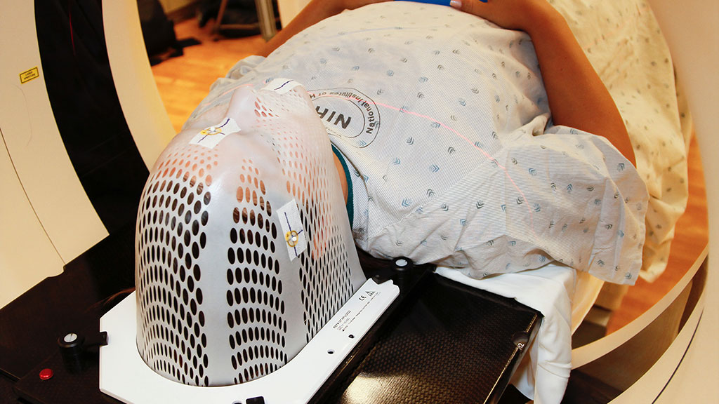
The technique helps analyse the effects of the coronavirus on patientsCT scans are one of the most effective ways to see inside the body
Computerized tomography (CT) is one of the most effective medical tests for analysing the effects of many illnesses, including COVID-19, on the lungs. An international team led by the UOC has developed a new method that improves the quality of the images obtained from CT scans. The algorithm, which has been tested on simulated data, enables them to distinguish different body’s tissue types better and opens the door to lowering the doses of radiation to which patients are exposed during this type of test.
A window on the body's internal workings
Computed tomography, or CT, utilizes computational processes to combine many X-ray measurements taken from different angles of the body to produce tomographic images. This non-invasive procedure, which provides a three-dimensional reconstructed view of organs or tissues, allows physicians to see inside the target without cutting.
"[This technique] helps experts to determine the presence of a tumour, its exact location, size and spread. It can also be used to diagnose muscle and bone disorders, infection or blood clots, heart disease, lung nodules, and liver masses," said Mohammad Mahdi Dehshibi, a postdoctoral researcher at the UOC's Scene Understanding and Artificial Intelligence laboratory (SUNAI), of the UOC's Faculty of Computer Science, Multimedia and Telecommunications, of the Pattern Research Centre in Teheran, Iran. "This technology is among the most common imaging tools used to guide biopsy and radiation therapy as well as monitoring the effectiveness of treatments like cancer treatment, internal injuries, and bleeding detection."
However, computed tomography involves the risk of damaging the structure of DNA and, subsequently, cancer due to the exposure of the body to high-dose X-rays. For example, in the course of a head CT scan, a person receives a dose of radiation equivalent to the total amount they are usually exposed to in 243 days of normal living.
A new algorithm to reduce radiation
In a search to reduce this radiation, the team led by Dehshibi has developed a new post-processing algorithm which increases the quality of reconstructed CT images. While conventional CT methods pick up only a part of the X-ray energy spectrum, the researchers tested a broader energy range, divided into intervals, to reach higher contrast. After testing it on constructed data using GATE/GEANT4 simulation software, they found that the algorithm enhances the quality of the images while reducing noise, which enables better discrimination between different types of tissue with lower doses of X-rays, according to their findings published in the Journal of Information Processing.
"Distinguishing between two different tissues (either normal or abnormal ones) in the same region is critical for physicians or radiologists to plan for further treatments, where this decision is dealing with the patients' lives," he said. "Having better tissue discrimination increases the success rate of the medicine's plan." The new method increases the capacity to distinguish between tissues by 60% in simulations compared to conventional CT.
"Our viewpoint was proposing a post-processing approach that does not need a substantial hardware reconfiguration and gives more freedom to imaging scientists for further exploration," Dehshibi said. "We hope that the findings of this study are later examined in the clinical setting to reduce the radioactive effect of irradiating with X-ray."
Related article
Gholami, N.; Dehshibi, M. M.; Adamatzky, A.; Rueda-Toicen, A.; Zenil, H.; Fazlali, M.; Masip, D. A Novel Method for Reconstructing CT Images in GATE/GEANT4 with Application in Medical Imaging: A Complexity Analysis Approach. Journal of Information Processing 2020:28, 161. Doi: https://doi.org/10.2197/ipsjjip.28.161
UOC R&I
The UOC’s research and innovation contributes to solving the challenges facing the global societies of the 21st century by studying ICTs’ interactions with human activity, with a specific focus on e-learning and e-health. Over 400 researchers and 48 research groups work among the University’s seven faculties and three research centres: the IN3, the eLearn Center and the eHealth Center.
The United Nations 2030 Agenda’s Sustainable Development Goals and open knowledge provide strategic pillars on which the UOC’s teaching, research and innovation are built. More information: research.uoc.edu.
Experts UOC
Press contact
-
Editorial department